- [课程通道] Gene Interference and Cat Allergen Silencing Research Project
- [课程通道]
- [科学少年] 2025-10-05
- [科学少年] 2025-10-02
- [科学少年] 2025-10-01
My Journey to the World of Nano medicine 爱博物 2025-10-01
【Preface】
In the summer of 2023, Arthur, a student from Tsinghua University High School Guanghua, participated in the Nano-drug Delivery Research Project organized by IBOWU · JSR. This article is his submission reflecting on and summarizing the project, now shared with everyone. We also welcome more JSR students to contribute and share the wisdom and joy that science brings you.
My Journey to the World of Nano-medicine
By Arthur
This was my brief yet eye-opening experience in the summer nano-medicine project. During the program, I followed professors, learning to synthesize nano-lipid carriers for the effective delivery of siRNA drugs into cancer cells. I witnessed the power of nanotechnology in modern medicine and gained a deeper understanding of the allure of scientific research. I invite you to explore this amazing microscopic world with me!
Cancer is one of the hardest diseases to treat, and many people, unfortunately, die from it. For instance, in China, about three million people lose their lives to cancer every year. The biggest challenge is there's no effective drug to treatment. During a week-long summer program with JSA (iBowu, Juvenile Science Academy), we studied how tiny particles (nano-particles) can help send medicine straight into cancer cells.
In our experiment, we utilized siRNA technology. Imagine our cells have little instruction books (DNA, and then RNA) inside them, telling them what to do. Sometimes, a page in that book might have wrong instructions that can make us sick (RNA of cancer cells). siRNA technology is like using a special eraser (siRNA drug) to remove that wrong page so the cell doesn't follow those bad instructions. This can help stop diseases. We combined this drug with nanoparticle liposomes, which helps get the drug into the cell. This method is beneficial because it works well in the body and is gentler on the cells
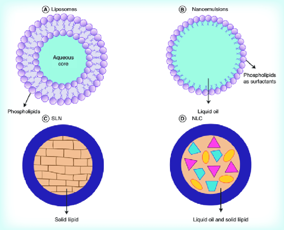
Figure 1. Schematic drawing of nanoparticle liposomes. Figure taken from open access publication: Chia Lang Fang et al., Delivery and targeting of nanoparticles into hair follicles, Therapeutic Delivery5(9):991 (2014). DOI: 10.4155/tde.14.61
The entire experiment was broken down into 8 steps, so it's easier to understand:
1. Hela Cell resuscitation We woke up some HeLa cells from liquid nitrogen, which kept the cells live but inactive at -78 degrees. We also gave HeLa cells fresh nutrient, or what scientists call a '‘culture solution”. We performed centrifugation to collect all the cells and move them to a new growing environment, and finally we observed through a microscope to determine if the cells were healthy and strong. In the experiment, we need to extract the cells gently and precisely, which is a big challenge.

Figure 2. HeLa cell resuscitation
2. Preparation of nanoparticles. We synthesized nanoparticles in special chemical solutions. Later, they will work like cars to carry the drugs into cancer cells. When making these nanoparticles, it is important to obey the order of adding each chemical into the solution. Adding them in a wrong order will lead to failure of synthesis.
3. HeLa Cell medium changing. We had to move the HeLa cells to new spots and give them more fresh nutrients so they can keep growing. We had to be very careful not to hurt the cells
4. Make siRNA medicine. At the same time, siRNA drugs can be made by adding previously designed pieces of RNA into a special chemical solution. The scientific jargon for it is “buffer”. Sufficient and gentle mixing is the key to success when making siRNA drugs.
5. Passage and transfection of HeLa cells. We then carried out the passage and transfection of Hela cells, that is to say, we let the cells grow their next generations to obtain more cells to work with. When adding cells to the plate, we had to be careful not to make foams. Meanwhile, we took care not to damage the cells while removing the medium.
6. Observe gene transfection efficiency. After passage and transfection, we tested the transfection rate of cells by observing and counting the live cells under a high resolution microscope.
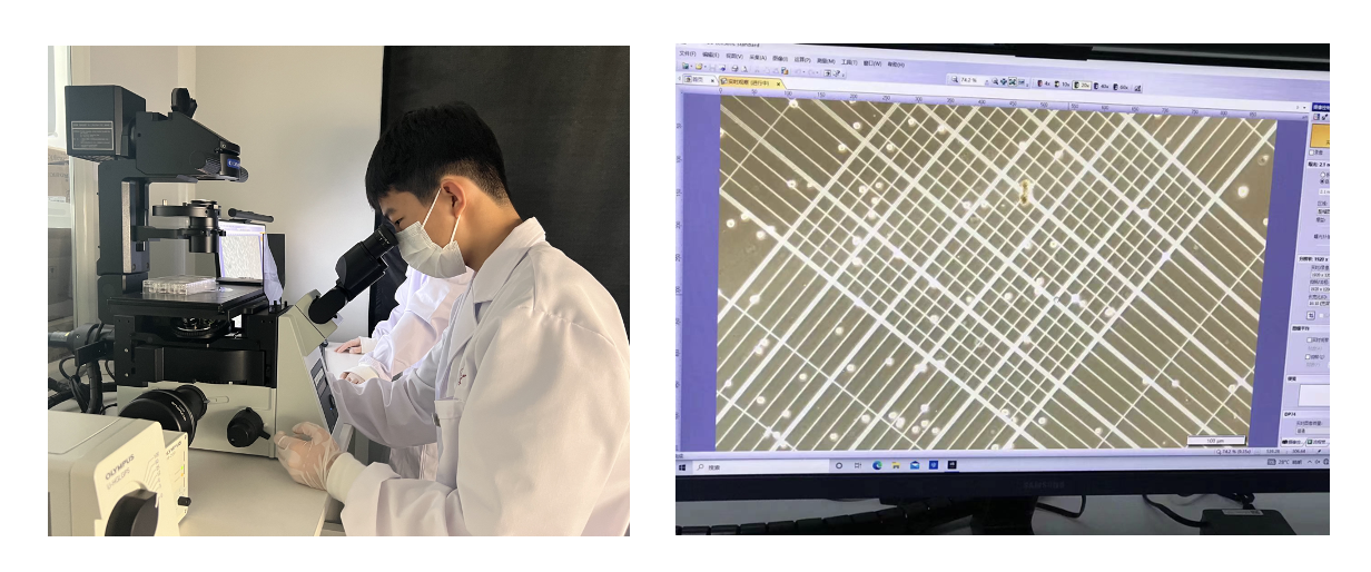
Figure 3. Observing cell passage and transfection efficiency under a microscope.
7. Test nanomedicine’s toxicity. All drugs can be toxicity to normal cells, and we needed to study how toxic they are. We added empty nanoparticles and nanomedicines that has siRNA drugs to healthy cells. The toxicity can be measured by counting how many cells can survive.
8. In vivo experiment in mice. Finally, we injected the drug into mice through the tail vein to reach the target organ through the bloodstream.
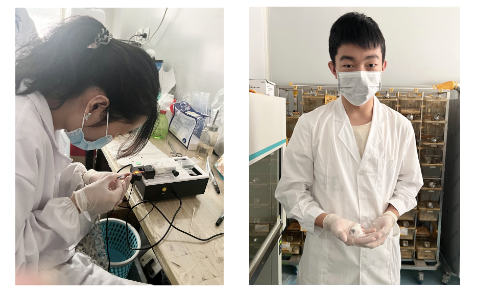
Figure 4. Injecting nanomedicine into the mouse with the help of a research teacher.
We examine the results of the experiment using many cutting-edge methods, such as Atomic Force Microscopy (AFM) and Flowcytometry.
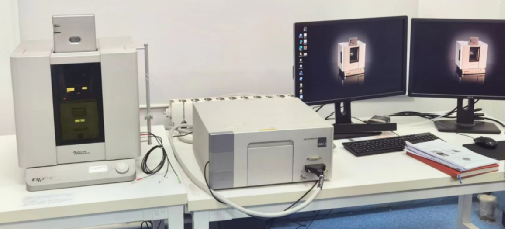
Figure 5. Atomic Force Microscope we used in our experiment.
This is what an empty nanoparticle looks like under AFM: tiny, only 20 nanometers in height.
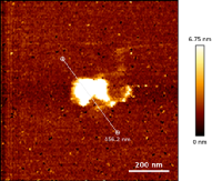
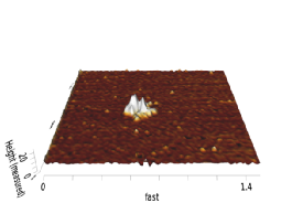
Figure 6. Observing empty nanoparticles under the Atomic Force Microscope
This is what nanoparticles containing siRNA look like under AFM, and it's bigger than last one.
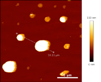
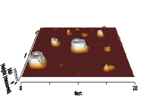
Figure 7. Observing nanomedicines under the Atomic Force Microscope
The next two pictures show the cells under a fluorescence microscope. The one on the left shows cells without adding the drug, and the one on the right shows cells 24 hours after adding drug. Brighter color means there are more living cells, and it shows our drug is effective in killing the cancer cell, i.e., HeLa cells.
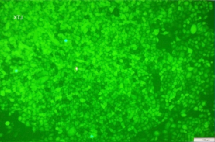
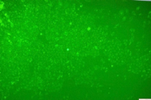
Figure 8. Observing cells with and without adding siRNA drugs under the fluorescence microscope
In the next few figures, we can count exactly how many cells are alive, using a special experiment device called flowcytometry.
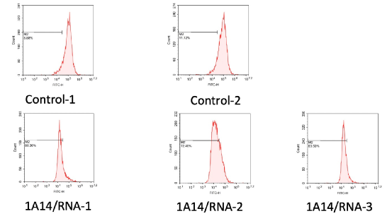
Figure 9. Counting the fluorescence using flowcytometry.
We combined the above data and compiled two bar charts to show a more clear comparison between adding and not adding drugs to the HeLa cells.
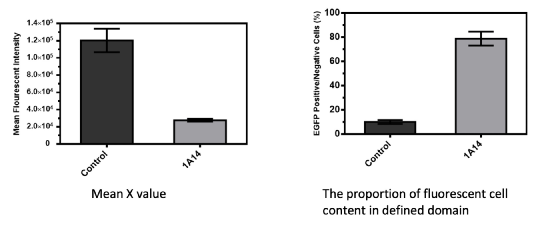
Figure 10. Counting the fluorescence using flowcytometry.
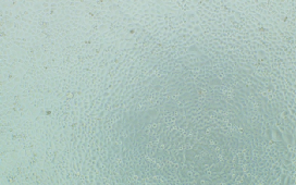
The next two pictures are what the cells look like under the microscope, and we can see that the cells on the left with RRM2 are larger, while the cells on the right without RRM2 are more.
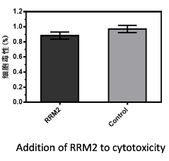
After testing the toxicity of these two groups by zymography we obtained a bar graph like this one. We can see that there is less toxicity in cells with added RRM2
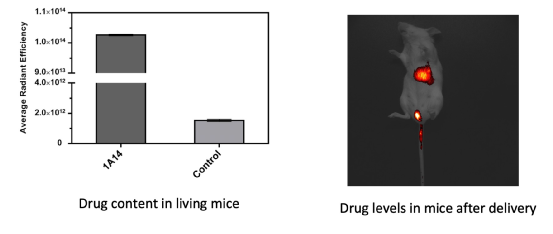
The last is the mouse in vivo experiment, where we use mice to simulate humans to test nanomedicine effectiveness. The figure on the right is the distribution of the drug in the mice. As you can see, there is still some drug residue in the tail because of irregular operation in the injection.
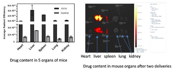
The main bar of our medications is found in the mouse liver.
There are still many shortcomings in our experiment. Our experiment is a proof-of-concept experiment, so few variables are discussed. This needs to be improved in the future.
For more information, please contact an IBOWU Course Advisor:
Ms. Zhang: 13910908618 (WeChat ID same as phone number)
Ms. Zha: 13910312020 (WeChat ID same as phone number)
About IBOWU · JSR
JSR™ is China's first junior science academy initiated by non-governmental forces. Launched by IBOWU, a well-known platform in China for providing scientific research and academic projects for youth, JSR aims to offer high-quality scientific practice platforms for Chinese teenagers.
JSR has received investment from BGI Group, TAL Education Group, and Poly Group. Leveraging strong scientific and capital backing, JSR has gathered dozens of multi-disciplinary frontline scientists from around the globe and established joint laboratory resources spanning numerous universities and research institutions in countries including China, the USA, Denmark, Norway, Singapore, and Australia. JSR is committed to providing students with high-end STEM advanced academic programs and elite training plans, striving to cultivate the scientific spirit among Chinese youth and contribute to the reserve of scientific and technological talent for China and the world.


